|
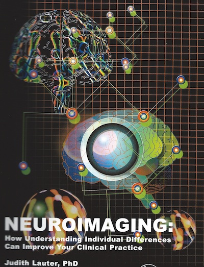
Neuroimaging: How understanding individual differences can improve your clinical practice
© American Speech-Language-Hearing Association, 2002 transferred to Judith Lauter, 2005 3-hr video of powerpoint presentation, plus 103-page manual (8.5" x 11") including all slides plus a set of article reprints publisher: American Speech-Language-Hearing Association, Rockville MD available on special order from the author (use email icon top right of this page) ***** Excerpts below include:
Following the excerpts is a 2002 review by Ray D. Kent PhD |
ABSTRACT
One of the most striking features of human beings is their dramatic degree of individual difference -- just as with fingerprints and zebra stripe patterns, each individual seems to be unique. In everyday life such diversity may be invigorating, but in clinical practice, it can lead to confusion in diagnosis, management, and predictions regarding outcome. New noninvasive techniques for brain imaging and monitoring promise revolutionary new insights into the neurobiological bases of individual differences, as well as new approaches to grouping, that may help resolve such confusions. This video conference will discuss a new perspective on individual differences by providing: 1) an image sampler illustrating individually-specific results collected with a variety of noninvasive methods; 2) a brief description of procedures and results using a new test battery designed for the study of individual differences, and based on relatively inexpensive technology well within the means of many clinicians and researchers in speech and hearing; and 3) discussion of a new brain model which has grown out of this type of testing, offering a “big picture” approach to the neural bases of individual differences as well as the origins of categories of characteristics, such as communication disorders.
OUTLINE
General outline
I. Introduction: Individual differences -- where do we find them?
II. Brains & zebras: Individual differences in brain imaging results
III. The AXS Battery: Capabilities for “neural fingerprinting” in your own clinic
IV. The Trimodal Model of Brain Organization: A new perspective on the why and how of individual differences
Expanded outline
I. Introduction: Individual differences: Where do we find them?
A. Human diversity in everyday life
1. Most familiar feature of humans
2. A biological commonplace
B. The exasperation of differences in the clinic
1. Diagnosis: To lump or to split?
2. Recommendations: The same dose for all?
3. Predicting and assessing outcome: “Snake oil” or -- hidden differences?
II. Brains & zebras: Individual differences in brain imaging results
A. Introduction
1. Old windows and new
2. Comparative overview of noninvasive methods
B. Sampler of individual results in brain anatomy
1. Computed Tomography (CT)
2. Magnetic Resonance Imaging (MRI)
C. Sampler of individual results in brain physiology (methods based on brain electricity)
1. Evoked Potentials (EPs)
2. quantitative Electroencephalography (qEEG)
3. Magnetoencephalography (MEG)
D. Sampler of individual results in brain physiology (methods based on other features)
1. Single-Photon-Emission Computed Tomography (SPECT)
2. Positron Emission Tomography (PET)
3. Functional Magnetic Resonance Imaging (fMRI)
4. Event Related Optical Signals (EROS)
III. The AXS Battery: Capabilities for “neural fingerprinting” in your own clinic
A. Overview of the AXS Battery
1. Designing a test battery that is both powerful & practical
2. Philosophy behind the AXS Battery
B. Physiology at the periphery: Otoacoustic emissions (OAEs)
1. The OAEs protocol -- background and procedures
2. Sample data (ear comparisons, effects of Ritalin, etc.)
C. A new window on the brainstem: REPs/ABR
1. The REPs protocol -- background and procedures
2. Sample data (staging brainstem development, effects of nicotine, etc.)
D. Top of the auditory world: qEEG in auditory cortex
1.The qEEG protocol -- background and procedures
2. Sample data (brain asymmetries, effects of Ritalin, etc.)
E. Putting it all together: The auditory system as microcosm
1. Co-variances linking ear, brainstem, and cortex
2. “Central neurolaryngology:” Exploring the neural bases of voice and speech
3. Practical applications in cardiology, reading, documenting outcome, etc.
IV. The Trimodal Model of Brain Organization: A new perspective on the why and how of individual differences
A. Individual differences and similarities: The history of human typology
1. Old systems from the Greeks to astrology
2. Body types and hormotypes
3. New sources of information: psychoneuroimmunoendocrinology
B. Bases of the Trimodal Model #1 -- Sex hormones and brain organization
1. Sources and actions of sex hormones
2. The schedule of testosterone exposure in humans
3. The Trimodal prediction: A spectrum of dosage from zero to high
C. Bases of the Trimodal Model #2 -- Functional asymmetries
1. Old approaches to functional asymmetries
2. The EPIC model and analogues in sensory & motor systems
3. Importance of functional asymmetries for IDs in personality, skills, health
D. Tenets of the Trimodal Model: Causes, continuua, capabilities
E. Implications of the Trimodal Model: Three varieties of human brain and their features
F. Applications of the Trimodal Model: Example = reading & learning skills
COURSE DESCRIPTORS
This video conference will:
o review the potential of currently available methods for noninvasive studies of individual differences in human brain and behavior, including all aspects of communication, whether hearing, speech or language
o identify and explain a new, relatively inexpensive test battery that brings sophisticated studies of this type within the range of many clinicians and researchers, even those with limited funding
o describe a new model of the brain based on brain plasticity during prenatal development with far-reaching implications for many features of health and behavior, including propensity for different types of disorders as well as individually differential capacity for functional recovery from injury
o predict the ways in which advances in understanding the neural bases of individual differences could impact on diagnostics, intervention, and outcome assessment in a wide variety of clinical populations, including hyperactivity, swallowing problems, autism, dyslexia, stuttering, Parkinson’s Disease, Alzheimer’s Disease, tinnitus, hyperacusis, and central auditory processing disorder
LEARNER OUTCOMES
After this video conference, audience members will be able to:
o consider the crucial importance which information on individual differences promises for our understanding of behaviors related to communication, whether focused on hearing, speech, or language
o analyze how their own professional and/or scientific activities could be enhanced through use of a new approach to framing test batteries using technology already available in many communication disorder clinics
o identify the “brain type” of individuals and consider how this can “explain” clusters of features involving personality, health, and communication ability, observed in the clinic and in everyday behavior
REVIEW BY DR. RAY D. KENT
(originally published in Tejas vol XXVI, p. 48, 2002; permission to use on author's website granted by Dr. Kent)
Professor Judith Lauter is highly experienced with a variety of neuroimaging methods used to examine brain functions underlying speech,language, and hearing. She has become an articulate and inspiring advocate for the application of these techniques to communication sciences and disorders, almost singlehandedly defining and promoting the emerging discipline of communication neuroscience. This self-study product consists of two videotapes and a companion workbook that reflect very well on Dr. Lauter's considerable expertise and her vision for the marriage of neuroimaging and communication disorders. These materials should be valued both by those who are new to these techniques (and would like a general introduction) and those who are more experienced (and would like to see new theories and applications). The tapes and workbook are divided into three major parts that follow a brief introduction: (1) Brains and Zebras: Individual Differences in Brain Imaging Results; (2) The AXS Battery: "Neurological Fingerprinting" in Your Own Clinic; and (3) The Trimodal Model of Brain Organization. These sections fit well together in developing the major thesis that individual differences are readily apparent through brain imaging and monitoring, that these differences can be understood through neurobiological principles, and that these differences have direct and significant clinical applications.
While briskly and clearly explaining the major neuroimaging techniques (MRI, EPs, REPs, qEEG, MEG, PET, fMRI, and EROS), Lauter presents pervasive evidence of individual differences revealed by these methods, thereby setting the stage for the following sections. AXS is an acronym for the Auditory Cross-Section Test Battery, which consists of three kinds of tests: behavioral background (e.g., audiometry), a three-level physiological profile (otoacoustic emissions, repeated evoked potentials/auditory brainstem response, and quantitative electroencephalography), and an array of complementary tests (to mention just a few, eye movements, speech acoustics, cardiac function). Use of the AXS Battery is illustrated by a variety of materials, including photographs, schematic diagrams, chart recordings, and data plots -- all of which justify the assertion that a coordinated test battery must integrate the examination of behavior, peripheral physics and physiology, and the central nervous system. The final section addresses the Trimodal Model of Brain Organization, which recognizes the interactions of genetics, environment and sex hormones to yield functional asymmetries in the brain underlying the three "modes" of left-favoring "focal," symmetrical "middle," and right-favoring "polytropic." Clinical implications are discussed with respect to a reading/learning skills pyramid.
When I first saw the term "trimodal," I was reminded of other tripartite descriptions of brain function, such as Paul MacLean's triune brain and A.R. Luria's three-part division of neural functions. But Lauter's concept is novel, and it holds intriguing potential for understanding human behavior and a variety of communication and learning disorders. Although the work is ambitious in its grasp, the basic concepts are explained quite simply, making them understandable to a wide audience. Lauter has been indefatigable in arguing that more attention must be paid to individual differences, including their biological and environmental origins, and their implications for clinical assessment and intervention. The recent literature gives support to her efforts. It has been reported that, at the population level, there is a graded continuum between left and right hemispheric language lateralization (Knecht et al., 2002), and this principle may account for some of the individual variability in response to unilateral brain lesions. Several reports show dissimilar patterns of lateralization between males and females, although different opinions have been expressed on the origin, nature, and significance of these differences (Amunts et al., 1999; Harasty et al., 1997; Kansaku & Kitazawa, 2001; Robichon et al., 1999; Vikingstad et al., 2000). Lauter has shown the way to a vein that may be rich with scientific and clinical ore. Her trimodal model is worthy of test, and, even it it should not turn out to be completely accurate, its examination will no doubt led to new understanding, and perhaps new horizons in biobehavioral science. She should be commended for her efforts to weld clinical practice and neurobehavioral science into a complementary and mutually productive connection.
The workbook includes 91 pages of slides, an 11-page annotated bibliography, an appendix of four reprints, a section of "next steps" (advice on reading the literature, using available resources, and learning new techniques), and a set of 25 self-study questions. The videotapes amplify the materials found in the workbook and also give a greater cohesion to the information. They are of good visual and auditory quality. The workbook is valuable because it helps to crystallize the ideas as "hard copy," and it includes extended discussions of the central concepts in the form of the four reprinted articles. The videotapes and workbook are an effective combination, inviting the viewer/reader to share in Lauter's experiences and creative thinking.
References
Amunts, K., Schleicher, A., Burgei, U., Mohlberg, H., Uylings, H.B.M., & Ziles, K. (1999). Broca's region revisited: Cytoarchitecture and intersubject variability. Journal of Comparative Neurology, 412, 319-341.
Harasty, J., Double, K.L., Halliday, G.M., Krill, I.J., & McRitchie, D.A. (1997). Language-associated cortical regions are proportionally larger in the female brain. Archives of Neurology, 54, 171-176.
Hausmann, M., Behrendt-Korbitz, S., Kautz, H., Lamm, C., Radelt, F., & Gunturkun, O. (1998). Sex differences in oral asymmetries during word repetition. Neuropsychologia, 36, 1397-1402.
Kansaku, K., & Kitazawa, S. (2001). Imaging studies on sex differences in the lateralization of language. Neuroscience Research, 41, 333-337.
Knecht, S., Floel, A., Drager, B., Breitenstein, C., Sommer, J., Henningsen, H., Ringelstein, E.B., & Pascual-Leone, A. (2002). Degree of language lateralization determines susceptibility to unilateral brain lesions. Nature Neuroscience, 5, 695-699,
Robichon, R., Giraud, K., Berbon, M., & Habib, M. (1999). Sexual dimorphism in anterior speech regions: An MRI study of cortical asymmetry and callosal size. Brain & Cognition, 40, 241-246.
Vikingstad, E.M., George, K.P., Johnson, A.F., & Cao, Y. (2000). Cortical language lateralization in right handed normal subjects using functional magnetic resonance imaging. Journal of the Neurological Sciences, 175, 17-27.
-- Ray D. Kent, PhD
Department of Communicative Disorders
University of Wisconsin-Madison
Copyright © 2023 Judith L. Lauter

